www.ch-thuer.ch Deutsch


Instability of the shoulder or shoulder girdle
 
 
Trauma-related instability of the shoulder
A dislocated shoulder is a common injury and is usually caused by falling onto the shoulder or outstretched arm. It is a typical sports-related injury, and one that particularly affects adolescents. In many cases, the humeral head can be replaced in its socket at the site of the accident. Occasionally, however, the patient will need to be taken to hospital to have the dislocation reduced under an appropriate anaesthetic. A shoulder dislocation can cause permanent damage to the joint, which may mean that the shoulder remains unstable and that the humeral head may pop out of its socket again when certain movements are carried out. Bone splintering at the edge of the socket or bone depressions on the head are possible, as are tear injuries of the limbus (edge of the joint meniscus) and associated ligaments. Arthro-MRI is the ideal method for diagnosing these injured structures. There is a rule that the younger a patient is when the first dislocation occurs, the greater the likelihood that chronic instability will occur as a result of repeat dislocations. We refer to these cases as repetitive, post-traumatic shoulder dislocations or post-traumatic shoulder instability

Figure on left:
1. Limbal tear, bottom
2. Limbal tear, top
3. Biceps tendon anchor

Figure on right:
Cross-section through the limbus and ligament structures in the normal state and after dislocation

Treatment
The dislocated shoulder should be reduced as soon as possible, but without using force.

Various methods have been described and recommended. Most important of all is the need for gentle and careful reduction so as to avoid causing any additional structural injury during the reduction manoeuvre. Although the joint does not necessarily need to be immobilised after reduction, it is recommended until the pain subsides, which usually takes 3-7 days.

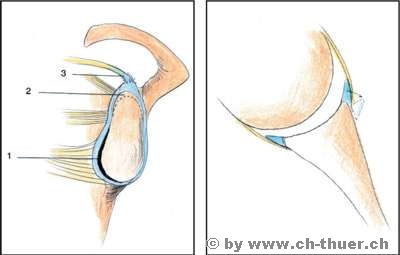
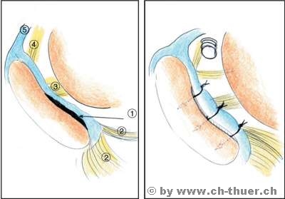
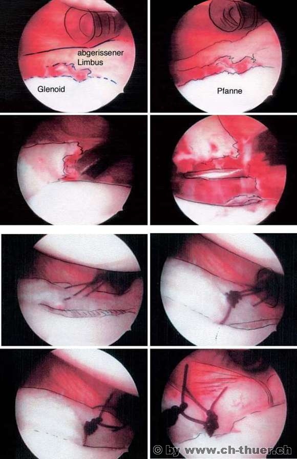
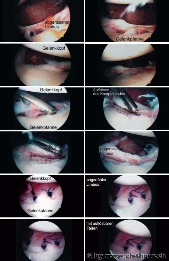
Repeated post-traumatic shoulder dislocations require surgical correction. Now that it is possible to use arthroscopy to manage these injuries and achieve very good outcomes, a number of surgeons and orthopaedists have adopted the practice of surgically treating first-time dislocations in young patients in order to avoid any extension of the joint damage in the event of repeat dislocation.

During the arthroscopic procedure, the torn meniscus and torn ligaments can be reattached to the anterior edge of the joint. This procedure spares the surrounding soft tissues and enables surgeons to rebuild the injured structures using dissolvable materials (absorbable sutures) so that the joint can be returned to its original state once the reconstruction has healed.

In some cases, however, open surgery will still be required, particularly for more extended injuries and repeated dislocations after a previous arthroscopic procedure.

Arthroscopic reconstruction

Figure on left:
1. Limbus detached from the edge of the socket
2. Lower ligament
3. Middle ligament
4. Upper ligament
5. Biceps tendon

Figure on right:
Following arthroscopic fixation of the meniscus to the anterior edge of the socket with 3 sutures

I now routinely treat cases of "normal shoulder instability" by arthroscopy. This is the ideal way of treating the structures of the joint from the inside using a minimally invasive procedure. Although, as mentioned above, this method has many advantages, it does involve the risk of the patient failing to comply with the immobilisation instructions because they are in minimal pain. This can in turn result in the formation of inferior scar tissue.

During the first three weeks after the operation, patients must wear an "Ortho-Gilet" (special immobilising sling) night and day and may only perform movements involving the elbow or forearm. External rotation of the arm is strictly forbidden. After three weeks, the shoulder need only be secured during the day with a normal sling. Patients still have to wear an Ortho-Gilet at night however. During the following three weeks, the patient can start performing active controlled movements of the shoulder: abduction (moving away) of the arm up to max. 90 degrees; external rotation of the arm up to max. 90 degrees.

Isometric exercises may also be started at this time. Pendulum exercises or exercises with weights are banned during the first 6 weeks. Only after 6 weeks are all securing devices removed and intensive physiotherapy started with appropriate movement exercises, which patients are also required to do by themselves at home. I always stress that passive stretching exercises - particularly exercises performed by a second individual - should not be performed during this phase of rehabilitation. Active strengthening of the dynamic shoulder stabilisers is crucial during this phase. Rehabilitation usually lasts 3-4 months. An even longer period with restriction of maximum external rotation is not only normal, but even desirable at this stage, because the scar tissue takes a few more months to become more elastic, after which full mobility is restored. A slow, gradual restoration of normal mobility tends to have a positive influence on the long-term outcome. Patients are usually are able to resume sporting activity and work at their pre-reconstruction level.